SPOTLIGHT: DiBio Imaging Facility
Pubblicato il: 23.02.2021 10:00
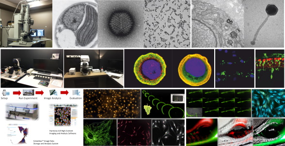
The facility manages advanced bioimaging equipment and provides technical and administrative management, equipment maintenance and upgrades, user training and support, laboratory and promotional activities:
Electron microscopy
The DiBioEM offers the following services:
preparation and observation of samples for conventional transmission electron microscopy - preparation and observation of macromolecules, cell fractions, nanoparticles, plastics and polymers - immunocytochemistry in pre and post embedding - technical and application advices - results interpretation - techniques update - equipment maintenance and upgrade - teaching activities - users training and tutorials -promotional activities.
Equipment
- TEM FEI Tecnai G2 with OSIS Veleta Camera
- Vitrobot FEI Mark III
- Ultramicrotomes: LKB Ultrotome III, LKB Ultrotome V, Reichert-Jung Ultracut 70170
- Sputter coater/glow discarge Edwards S150B
- Critical point dryer Polaron CDP7501
Optical and confocal microscopy
The different instruments cover a wide range of microscopy applications: analyses of fixed and in vivo samples as well as non-biological specimens employing a thermostated and CO2 chamber, DIC and phase contrast and different fluorescent filters (DAPI, FITC, Cy3, Cy5, Chlor, etc.)
Equipment
- Zeiss LSM 700 confocal microscope
- Leica Sp5 confocal microscope
- Leica MZ16
- Leica DMR
- Leica 5000B
- Leica DMI 4000B
Hight-Throughput Screening Facility
About us:
Since June 2019 the High Throughput Screening (HiTS@UniPD) facility at the Department of Biology, University of Padua, allows to perform medium/high throughput screens as well as small-scale research projects that can benefit from the use of our equipment and expertise in automation, assay development and data acquisition.
HiTS@UniPD MISSION
To provide screening services and access to state-of-the-art high throughput imaging technologies;
To offer the best support if you need to unbiasedly address life science questions.
Equipment and applications
-An Operetta high content Imaging system (Perkin- Elmer);
- Two automated liquid handling platforms;
- Chemical compounds and genome wide libraries;
- Automated nucleic acid purification platform.
Our technology is suitable for any type of sample, from 2D to 3D biological models including cell monolayers, isolated muscle fibers, parasites, moss/plants, spheroids, organoids and whole organisms (e.g. Zebrafish).
Your applications, your way:
• Fixed cell assays;
• Live cell experiments (as an example, FRET);
• Imaging of 3D cell models;
• Analysis of Complex cell models (e.g. o-culture systems, iPSCs, primary cell cultures);
• Drug discovery screens;
• Zebrafish imaging;
• Quantification of:
-Fluorescence intensity;
-Number of cells;
-Area of nucleus or cells;
-Mean intensity of nuclear and cytoplasm fluorescence;
-Organelles morphology;
-Analysis of cell differentiation, cell proliferation and migration.
Contact us for additional details:
DiBio Imaging Facility
Department of Biology,
Via U. Bassi, 58/B,
35131, Padova (PD) Italy
E-mail: imaging.biologia@unipd.it; hits.biologia@unipd.it;