SPOTLIGHT: Mouse embryos made with stem cells – an interview with Dr. Amadei
Pubblicato il: 29.09.2022 14:04
In the field of mammalian embryo development, mice are still the preferred model organism, but they are not perfect. First of all, they are very expensive to breed and maintain in a colony and secondly, quite a bit of time is required to genetically modify them to establish a novel transgenic mouse line. Since the discovery of embryonic stem cells, many researchers have tried to push them one way or the other to acquire the fate of any cell type of choice, or to recapitulate aspects of the developing embryo.
Here we interview Dr. Gianluca Amadei, of the “Cell Biology and Developmental Genetics” unit, expert in stem cell models of mouse embryogenesis, who recently joined DiBIO and is hosted by the research group led by Prof. Graziano Martello.
Amadei says "During my postdoctoral work in the group led by Prof. Zernicka-Goetz at the University of Cambridge, UK, my colleagues and I have found that we can go beyond capturing discreet aspects of embryonic development and actually model the entirety of the developing mouse embryo. To do so, we mixed together three types of mouse stem cells. Embryonic stem cells: they are the ones that can be a part of the actual embryo; the trophoblast stem cells: they can make tissue derivatives of the embryonic portion of the placenta; induced XEN cells: these are cells that were embryonic in origin, but that are manipulated to become cells like the ones of the primitive endoderm, and they will make differentiated derivatives such as the yolk sac. Once we mix these cells together and allow them to develop, over eight days they make structures that accomplish several impressive feats of development: they make an egg cylinder, complete gastrulation, undergo neurulation and the beginning of organogenesis. These structures, that we call ETiX-embryoids, not only do they look like real mouse embryos morphologically, but also transcriptionally they are remarkably similar.
What do I hope to achieve by making these structures? I hope that over time they will become more and more a versatile tool that can be used to model mouse embryonic development without resorting to actual mice. Of course, with any sort of artificial system, there are always potential improvements to be made, and trying to increase the amount of development these structures can recapitulate is always an exciting, albeit difficult, challenge. Here in the Dept. of Biology of the University of Padova I am keen to see what the system can and cannot do and use it to learn new and exciting biology. I hope that it will be a nice and useful complement to the ongoing research in the Department and that other groups will be interested in giving it a try!"
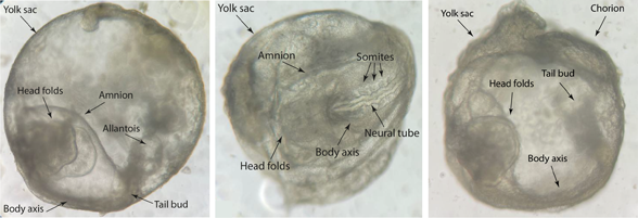
The picture shows brightfield images of ETiX-embryoids at seven days of development, which is closest to embryonic day 8.0 of natural mouse embryos. Left and right images show the side view, the centre image shows the ventral view. Features of the structures are labelled in each image. Images were captured by Gianluca Amadei.